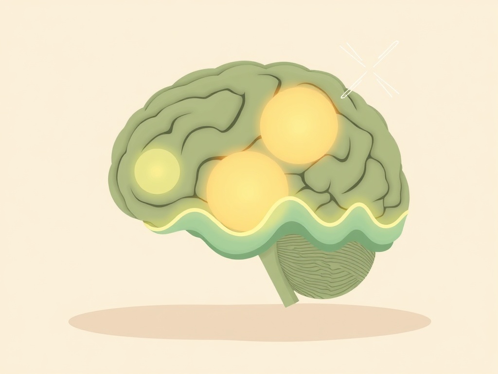
Social interaction difficulties may stem from disrupted neural scaffolding in an unexpected brain region. While autism spectrum disorders have traditionally been linked to cortical dysfunction, emerging evidence points to the cerebellum's critical role in orchestrating social behaviors through specialized protein networks that regulate neuronal firing patterns.
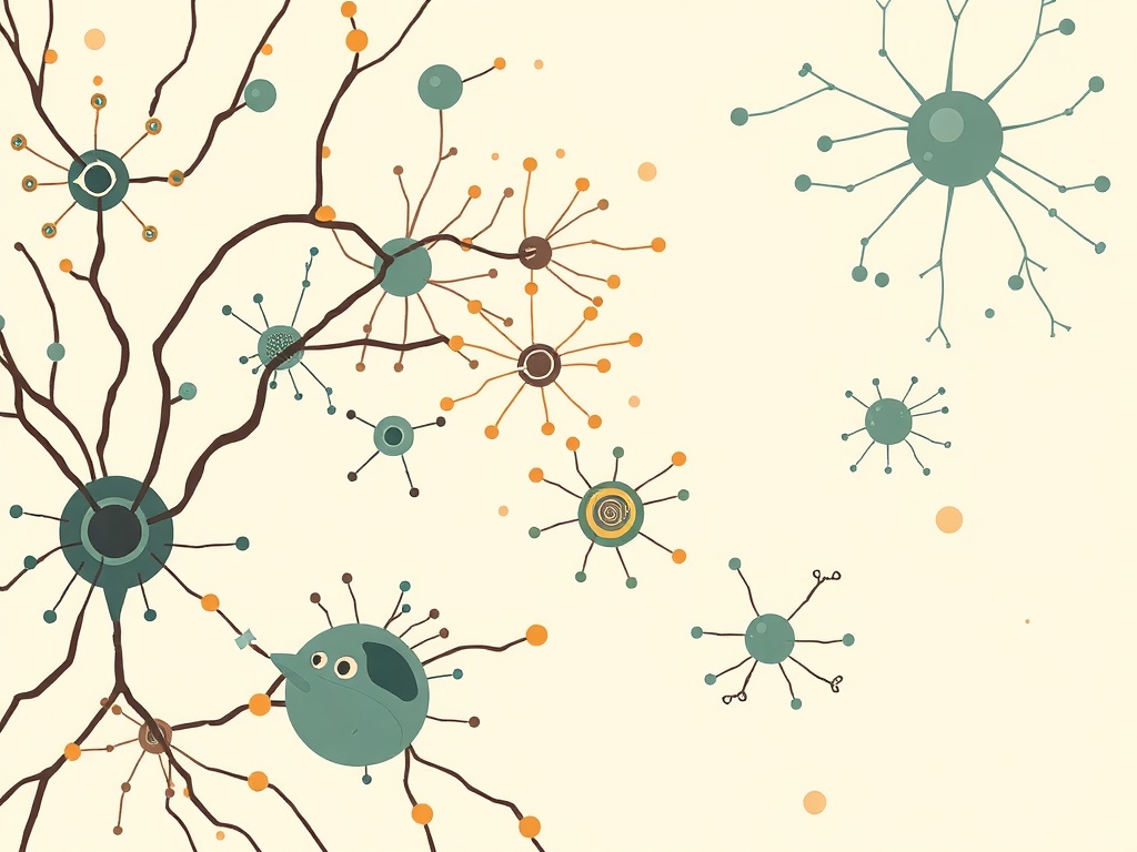
Researchers discovered that perineuronal nets—extracellular protein matrices that wrap around specific neurons—are significantly reduced in the cerebellar nuclei of autism-associated mouse models. When these protective nets were experimentally degraded using chondroitinase ABC enzyme, mice exhibited pronounced social interaction deficits. The treatment triggered hyperactivity in large glutamatergic neurons during social encounters, evidenced by elevated intracellular calcium dynamics and increased CREB1 phosphorylation. Additionally, the transcription factor ARNT2, which normally regulates neural activity, became abnormally elevated even without stimulation.
This finding challenges conventional understanding of autism neurobiology by implicating cerebellar dysfunction in social cognition. The cerebellum, traditionally viewed as a motor control center, appears to fine-tune social behaviors through precise regulation of neural circuit activity. Perineuronal nets act as molecular brakes, preventing excessive neuronal firing that could disrupt downstream social processing networks. Their degradation creates a cascade of hyperactivity extending to the red nucleus and ventromedial thalamic regions, suggesting cerebellar influence on distributed social brain networks.
While promising for understanding autism mechanisms, this mouse model research requires validation in human studies. The findings nonetheless suggest therapeutic approaches targeting cerebellar perineuronal net integrity could potentially address core social symptoms in autism spectrum disorders, representing a paradigm shift from cortex-focused interventions.