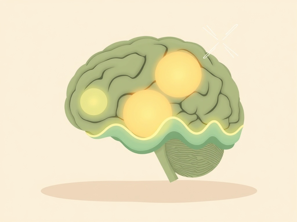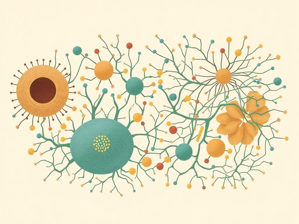
The molecular basis of social behavior may hinge on specialized protein scaffolds that wrap around specific brain neurons, potentially offering new therapeutic targets for autism spectrum disorders. These extracellular matrix structures, called perineuronal nets, appear to function as critical regulators of social interaction through their influence on cerebellar neural circuits.
Researchers discovered that mice with autism-related genetic modifications showed significantly reduced perineuronal net expression in cerebellar nuclei neurons. When these protein networks were experimentally disrupted using chondroitinase ABC enzyme, previously normal mice developed profound social interaction deficits. The cerebellar glutamatergic neurons showed increased activity during social behaviors, measured through calcium dynamics and CREB1 phosphorylation patterns. Additionally, the transcription factor ARNT2, which regulates neuronal activity, was elevated in both enzyme-treated and autism-model mice even without external stimulation.
This finding challenges traditional views of autism as primarily a cortical disorder by highlighting the cerebellum's central role in social cognition. The perineuronal nets appear to function as molecular brakes on neural activity, and their disruption leads to hyperexcitation that impairs social processing. The research connects cellular-level changes in protein scaffolding to complex behavioral outcomes through specific neural pathways extending from cerebellum to red nucleus and thalamic regions. While promising for understanding autism's neurobiological foundations, this mouse-based research requires validation in human studies. The work suggests potential therapeutic approaches targeting perineuronal net restoration, though such interventions remain years from clinical application.