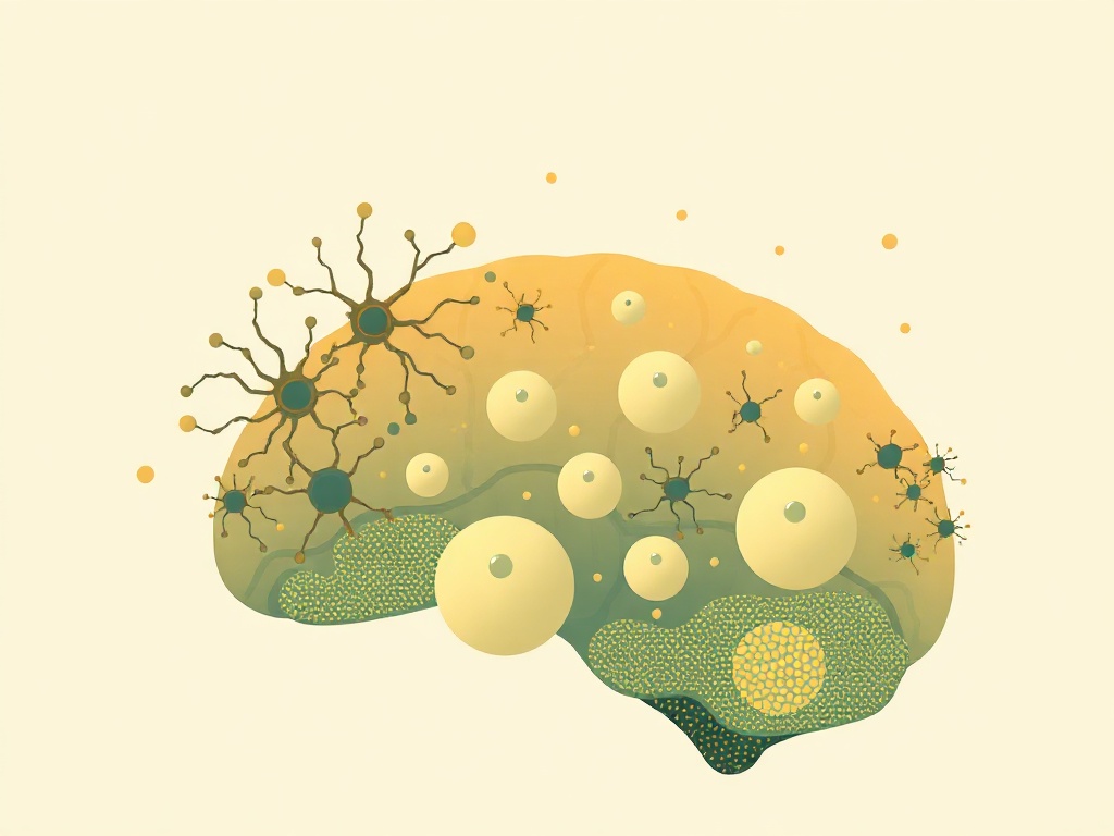
Understanding how the fetal brain constructs its neural architecture could reshape approaches to preventing developmental disorders and optimizing cognitive potential from the earliest stages of life. The transient subplate layer, which exists only during fetal development before dissolving, serves as crucial scaffolding for proper brain wiring but has remained poorly characterized in living humans.
Researchers used advanced T2-weighted MRI to map subplate development across different brain regions in human fetuses, revealing distinct growth trajectories and left-right asymmetries. The subplate showed marked regional variation in thickness and development timing, with some areas expanding rapidly while others remained stable. These patterns suggest the subplate's role extends beyond simple scaffolding to actively directing region-specific neural circuit formation.
This mapping represents a significant advance in developmental neuroscience, as previous subplate research relied heavily on post-mortem tissue analysis. The ability to track this ephemeral brain structure in living fetuses opens new possibilities for early detection of neurodevelopmental conditions. Since subplate abnormalities are linked to autism spectrum disorders, cerebral palsy, and schizophrenia, these MRI techniques could eventually enable prenatal screening for developmental vulnerabilities. However, the study's observational nature means causal relationships between subplate patterns and later outcomes remain unclear. The research also highlights how temporary fetal brain structures, despite their brief existence, may have lasting impacts on lifelong cognitive function and neurological health.