Understanding what makes a cell senescent — and distinguishing it from neighboring healthy tissue without chemical labels — has been one of cellular biology's most stubborn technical challenges. A new multimodal framework published in Nature Aging may substantially change how researchers detect, map, and ultimately target these aging-associated cells across tissues, with potential implications for longevity medicine and regenerative therapies.
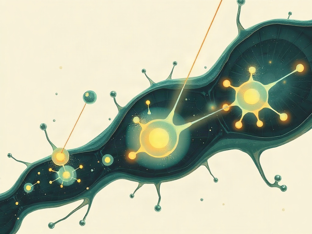
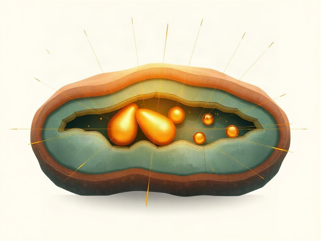
The work by Zhang, Chen, Monticolo, Sorrentino and colleagues integrates label-free Raman spectroscopy imaging with spatial transcriptomics and single-cell transcriptomics in a combined approach the team calls RamanOmics. By pairing the vibrational fingerprints of molecules — captured without staining or genetic modification — with gene expression data, the researchers were able to interrogate the biochemical architecture of senescent cells at high spatial resolution. Their central finding is a lipid-linked Raman spectral signature specific to p21-positive cells, the canonical marker of cell cycle arrest that defines many forms of cellular senescence. This signature was identified across both aging contexts and tissue repair scenarios, suggesting it may reflect a fundamental biochemical state rather than a context-specific artifact.
This finding carries several important dimensions worth contextualizing. The senescence field has long grappled with the absence of a single universal biomarker; p16, p21, SA-β-gal, and SASP panels each have limitations in specificity or detectability in vivo. A label-free optical signature that co-localizes with p21 expression could, in principle, enable non-destructive spatial mapping of senescent burden in tissue sections, and eventually inform drug target validation for senolytics or senomorphics. The lipid connection is biochemically plausible — dysregulated lipid metabolism is increasingly recognized as a feature of the senescent phenotype, potentially linked to lysosomal dysfunction and membrane remodeling. Key limitations include the current reliance on ex vivo tissue and the unknown translatability to live-tissue or clinical imaging contexts. As a methodology paper from a high-impact journal, this represents a potentially paradigm-shifting technical contribution rather than incremental progress.