Understanding why female reproductive aging precedes systemic aging by decades—and what cellular mechanisms drive that gap—has long been a missing piece in longevity biology. A high-resolution molecular map of the aging ovary could reshape how scientists conceptualize hormonal decline, cellular senescence, and the downstream effects on healthspan in women.
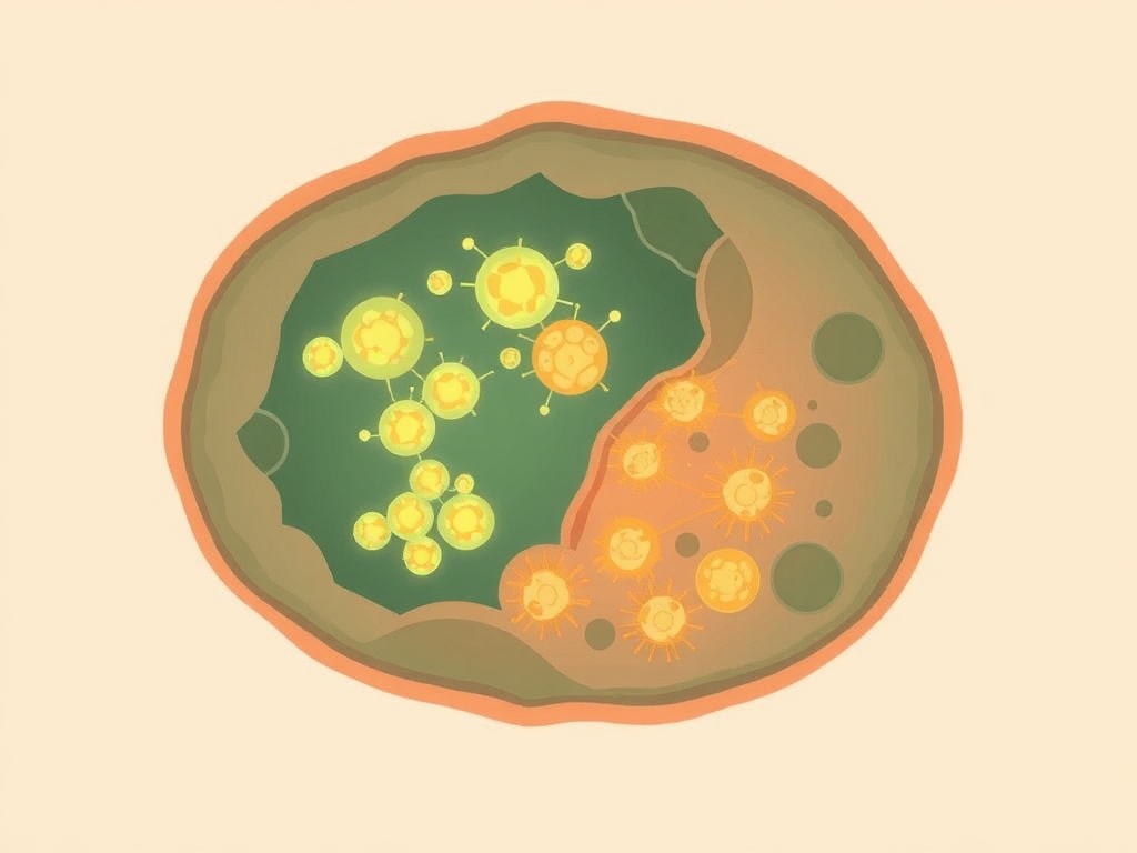
Published in PNAS, this study constructs a comprehensive single-cell transcriptomic atlas of the mouse ovary across precisely defined reproductive stages, including the transition into what the authors term the "estropausal" period—the murine analog of perimenopause. The atlas identifies ovary-specific senescent cell populations and characterizes their molecular signatures, revealing distinct transcriptional programs that differ from canonical senescence patterns described in other tissues. The researchers map cell-type-specific gene expression changes across granulosa cells, theca cells, stromal fibroblasts, and immune populations, providing a granular view of how cellular composition and signaling shift as reproductive capacity declines.
This work arrives at a moment when ovarian aging is receiving serious mechanistic attention as a lever for broader longevity intervention. Prior research established that senescent cells accumulate in aged ovaries and contribute to inflammation and follicle depletion, but lacked the single-cell resolution to distinguish which populations drive pathology versus which represent adaptive responses. By defining stage-specific molecular drivers, this atlas creates a reference framework that could accelerate target identification for senolytic or hormonal interventions. The mouse model is a meaningful limitation—human ovarian aging timelines and hormonal dynamics differ substantially—and single-cell RNA sequencing captures transcriptional state, not protein activity or causal directionality. Nevertheless, for a field that has historically relied on bulk tissue analyses, this level of cellular granularity represents a genuinely significant methodological advance. Its translational value will depend on validation in human ovarian tissue, but as a mechanistic foundation, the contribution is substantial.