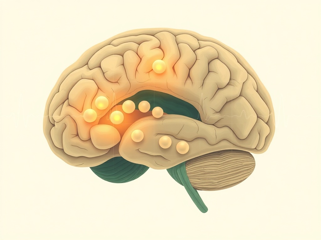
Sleep disruption often precedes cognitive decline in Alzheimer's disease by years, yet the biological machinery connecting amyloid accumulation to fragmented sleep has remained frustratingly opaque. New preclinical evidence now points to a specific cellular culprit — microglia, the brain's resident immune cells — and demonstrates that removing them rescues substantial sleep without touching amyloid burden itself. This reframing has real implications: it suggests sleep loss in early AD may be modifiable independent of plaque clearance.
Working with APP/PS1 transgenic mice at multiple disease stages, researchers combined EEG/EMG sleep recording with whole-brain light-sheet microscopy to map amyloid load and microglial density simultaneously. Amyloid plaques produced a reproducible loss of non-REM sleep that — crucially — did not escalate as plaque burden increased, suggesting a threshold rather than cumulative effect. More striking, microglial expansion spread well beyond plaque-adjacent zones into thalamocortical circuits and white matter tracts that regulate sleep-wake cycling, while the cortex showed signs of hyperexcitability and network desynchrony. When microglia were pharmacologically depleted via CSF1R inhibition, animals recovered more than two hours of daily sleep with no measurable change in amyloid levels, directly implicating neuroinflammatory signaling rather than plaque load per se.
This work sits at an important intersection: it challenges the implicit assumption that amyloid is the proximate driver of early AD symptoms, instead casting neuroinflammation as a semi-independent pathological layer. CSF1R inhibitors are already being investigated in other neurological contexts, so the translational pathway is not implausible. That said, mouse models of amyloid pathology have repeatedly shown species-specific sleep architecture differences, and APP/PS1 mice do not fully replicate the tau co-pathology or synaptic loss of human AD. The finding that normal aging alone eliminated sleep rebound capacity adds a meaningful confound for any human translation. Still, the identification of EEG desynchrony and microglial markers as presymptomatic biosignatures is an incremental but genuinely useful advance for early detection research.